Techniques

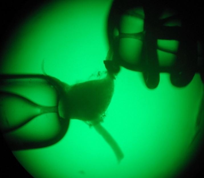
We were first to develop ‘Patch-seq’, the most advanced cellular profiling workflow widely used in neuroscience to classify neurons (Fuzik et al., Nature Biotechnology, 2016). Patch-seq takes advantage of the genetic tagging of cell types, their recording by slice electrophysiology (or even in vivo), intracellular biocytin loading for post-hoc neuroanatomy, and single-cell RNA-seq for molecular profiling.

Induced pluripotent stem cells (iPSCs) offer a powerful tool for our research because of their unique ability to differentiate into various cell types in the body including also neurons, microglial cells and astrocytes. Furthermore, by using CRISPR/Cas9 gene editing, we can generate isogenic iPSC lines with patient-specific mutations that are invaluable to study the disease mechanisms underlying, for example, altered brain lipid metabolism or neurotransmission.
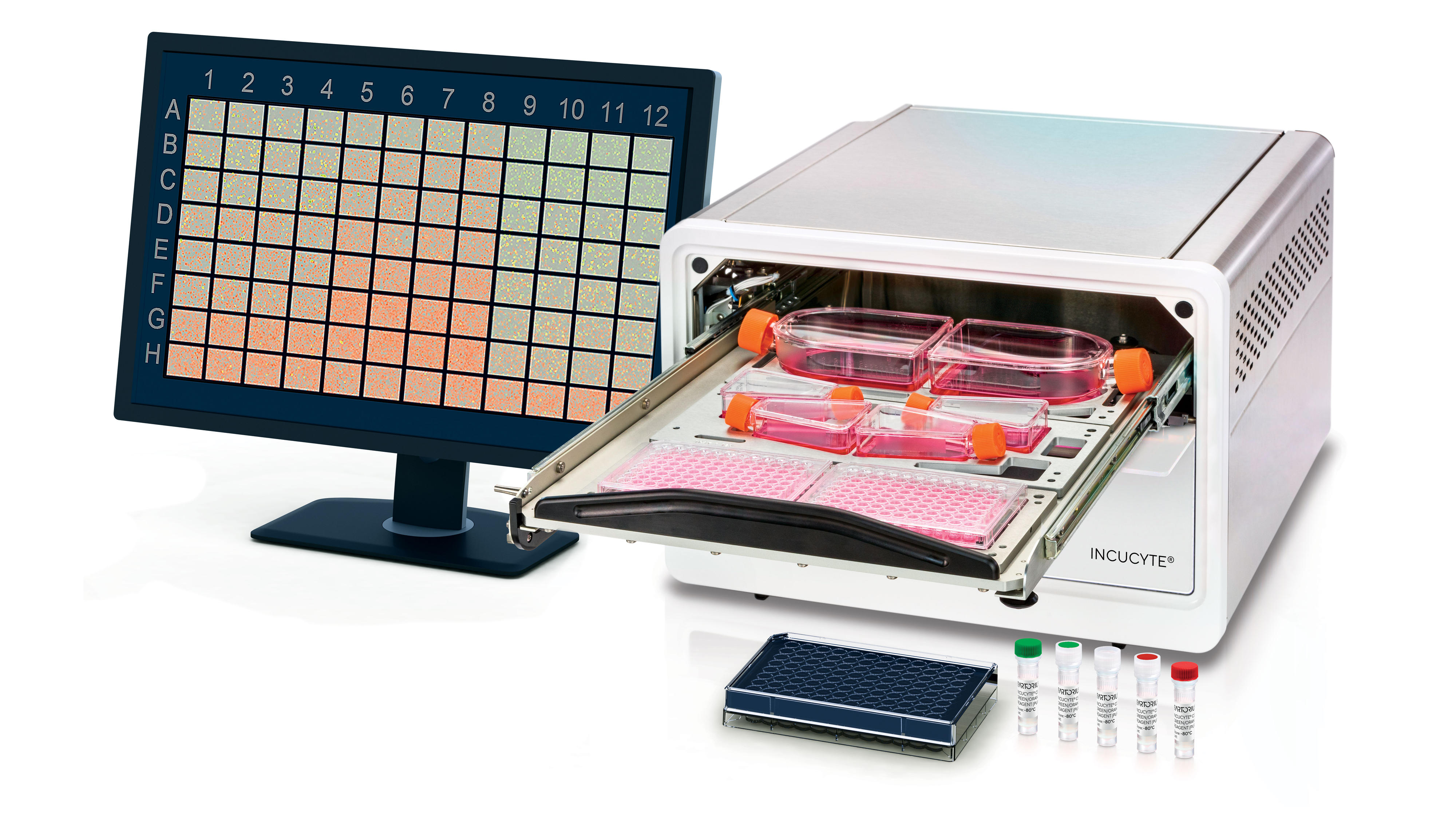
We have established the Sartorius IncuCyte SX5 multicolour live-cell imaging system, which allows high-throughput analyses of cell survival, migration, and differentiation or drug testing screens. This live-cell imaging system is also of particular utility for investigating microglial phagocytosis and neurotoxicity of lipids.

The Simoa technology represents a breakthrough in biomarker detection, allowing for ultra-sensitive quantification of proteins at femtomolar concentrations. This amplified sensitivity enables us to detect low-abundance biomarkers, like NfL, that are indicative of brain damage in blood and other body fluids. Access to the Simoa technology is enabled through our collaboration with Prof. Thomas Berger and Prof. Paulus Rommer (Institute of Neurology, MedlUniv Vienna).
For our biomarker research of plasma-derived cell-free DNA, we use EM-seq, a method available through the Sequencing Facility of MedUniv Vienna. EM-seq is a cutting-edge technology for analyzing DNA methylation patterns, which are essential markers for gene regulation and play a critical role in health and disease.

Our repertoire of molecular biology techniques includes CRISPR/Cas9 gene editing, analysing gene expression through RNA-seq and RT-qPCR, cell-free DNA isolation, FACS, magnetic-activated cell sorting (MACS) of different brain cell types, next to various other basic molecular biology techniques. For protein analysis, we are introducing the SimpleWestern system (Biotechne) as a quantitative alternative to conventional western blotting.
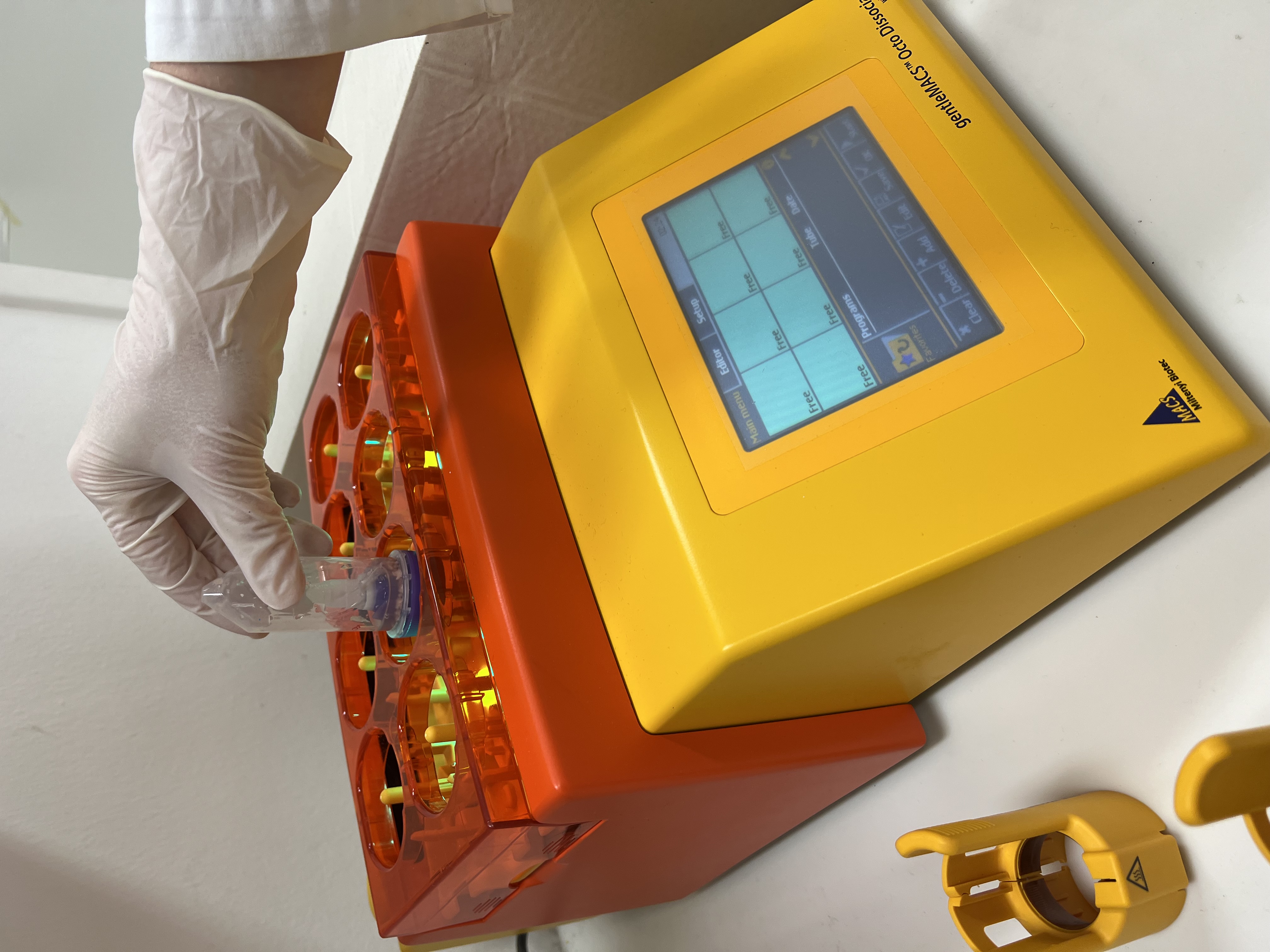
To isolate different brain cells, we use the gentleMACS™ Octo Dissociator to generate a single-cell brain suspension, followed by magnetic cell separation (MACS) targeting specific brain cell types. To determine cell purity and activation status, we proudly utilize our Guava® easyCyte flow cytometer, which is equipped with 3 different lasers.


