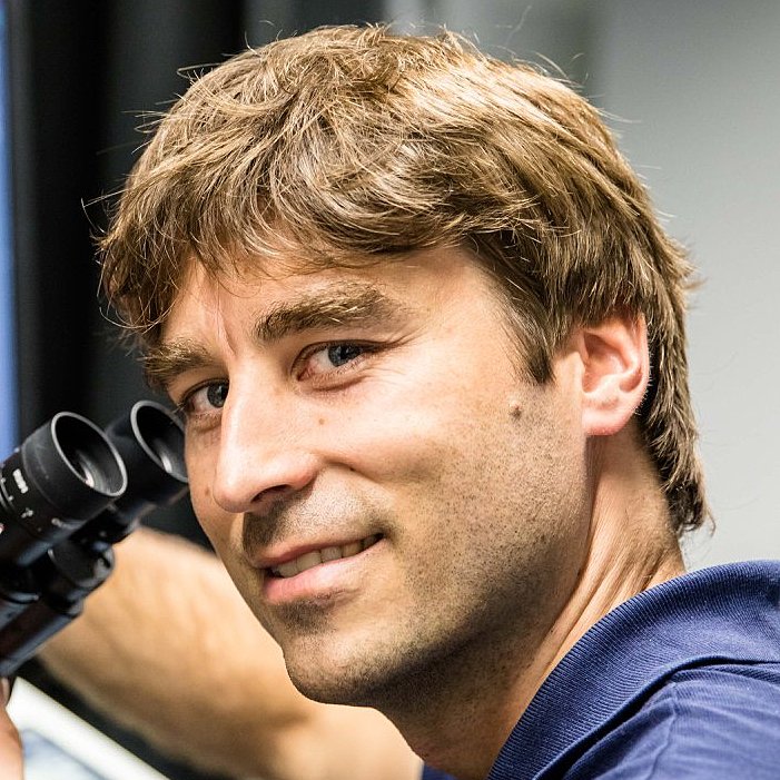
Prof. Dr. Johann Danzl

The Danzl lab focuses on the development of imaging technologies to extract new qualities of information from biological specimens with a strong focus on reconstructing brain tissue architecture. Recent advances include nanoscale reconstruction of living brain tissue (Velicky et al., 2023), a technology for analyzing brain tissue architecture across spatial scales (Michalska et al., 2023) and the development of light microscopy based connectomics (LICONN, Tavakoli et al., 2024). LICONN is the first technology beyond electron microscopy that enables dense, synapse level reconstruction of neuronal circuits, and opens up in-depth molecular characterization in connectomics. The group takes an approach across disciplines, with neuroscientists, biologists, physicists and computer scientists working together to address key gaps in biological measurement technology.
Focal points of interest
The Danzl group has a strong interest in reconstructing the functional architecture of neuronal circuits. Based on the LICONN technology, the group will integrate molecular characteristics into synapse-level circuit reconstruction, molecularly identifying cell types and states. The group is interested in deciphering how specific cell types, notably GABA cells, integrate in the circuit and how this mirrors molecularly and functionally defined subtypes. We want to investigate the principles of connectivity in GABA cells in rodent and human samples. Collaborating with groups at ISTA (P. Jonas, S. Siegert) and with the Medical University Vienna (K. Rössler, R. Höftberger), we have adapted our technologies to human brain tissue and, with G. Novarino, to cerebral organoids (Watson et al., 2024; Michalska et al., 2023, Velicky et al., 2023). We will refine technologies for reconstruction of human brain tissue at increased molecular information content and collaborate with P. Jonas to integrate structural and molecular information with functional characteristics obtained through electrophysiological recordings. Finally, we plan to take advantage of LICONN’s ability to analyze tissue architecture and connectivity across samples to address biological variability. This will allow defining differences in cellular abundance, distribution, connectivity and molecular characteristics across genotypes and in brain disease, including neurodevelopmental and neurodegenerative diseases.
Technical proficiency and instrumentation
The Danzl lab focuses on the development of novel technologies based on optical imaging and advancements in sample preparation. Directly integrating these with specifically tailored data analysis methods allows accessing information from biological systems that has been difficult to obtain otherwise. Expertise includes development and application of optical super-resolution technologies, including stimulated emission depletion (STED) and single-molecule methods. Furthermore, the group is proficient in the development of advanced sample preparation strategies, most notably expansion microscopy, as well as technologies to label specific molecules on protein and transcript levels. A major asset has been the group’s cross disciplinary approach, directly integrating computational analysis including deep-learning based methods into the technological developments. Recent examples include the development of LIONESS (Velicky et al., 2023), an integrated optical super-resolution/machine learning technology to reconstruct living brain tissue at nanoscale resolution. The CATS technology (Michalska et al., 2023) comprehensively visualizes brain tissue architecture by extracellular labeling compatible with chemical fixation and downstream super-resolution imaging. The group’s recent development of light microscopy based connectomics (LICONN, Tavakoli et al., 2023) is based on a specifically engineered expansion microscopy approach that provides the resolution, structural preservation, comprehensive contrast, and signal-to-noise ratio required for tracing even the thinnest of neuronal processes. Here, the sample is embedded in a swellable hydrogel and expanded, thus increasing effective resolution by moving cellular features further apart. With consecutive application of two swellable hydrogels and resulting 16-fold resolution increase, optical imaging with off-the-shelf optical microscopes achieves effective resolution of better than 20 nm, directly combined with highlighting of specific molecules.
The group has further expertise in development of volumetric spatial transcriptomics technologies (Agudelo et al., in preparation) to map brain tissue in 3D.
Aspirations for the next 5 years
The Danzl lab aims to develop the technologies to integrate information on cellular connectivity, molecular characteristics, and activity in the same cells and circuits, and to apply these technologies to map brain tissue in humans and rodents across health and disease. This will help establish disease state to tissue phenotype correlates, helping to gain mechanistic understanding, with potential implications for disease diagnostics and therapeutic approaches, both in brain and in other organ systems.
References
- Light-microscopy based dense connectomic reconstruction of mammalian brain tissue. Tavakoli MR, Lyudchik J, Januszewski M, Vistunou V, Agudelo-Duenas N, Vorlaufer J, Sommer C, Kreuzinger C, Oliveira B, Cenameri A, Novarino G, Jain V, Danzl JG Nature (May 2025) DOI: 10.1038/s41586-025-08985-1
- Imaging brain tissue architecture across millimeter to nanometer scales. Michalska J, Lyudchik J, Velicky P, Stefanickova H, Watson JF, Cenameri A, Sommer C, Amberg N, Venturino A, Roessler K, Czech T, Höftberger R, Siegert S, Novarino G, Jonas P, Danzl JG (2023) . Nature Biotechnology 42, 1051–1064.
- Dense 4D nanoscale reconstruction of living brain tissue. Velicky P, Miguel E, Michalska JM, Lyudchik J, Wei D, Lin Z, Watson JF, Troidl J, Beyer J, Ben-Simon Y, Sommer C, Jahr W, Cenameri A, Broichhagen J, Grant SGN, Jonas P, Novarino G, Pfister H, Bickel B, Danzl J (2023). Nature Methods 20, 1256–1265.
- Human hippocampal CA3 uses specific functional connectivity rules for efficient associative memory. J. F. Watson, V. Vargas-Barroso, R. J. Morse-Mora, A. Navas-Olive, M.R. Tavakoli, J.G. Danzl., M. Tomschik, K. Rössler, P. Jonas.
Cell (Dec 2024) DOI: 10.1016/j.cell.2024.11.022


