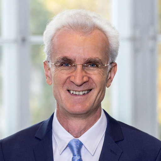
Univ.-Prof. Dr. Karl Rössler

Epilepsy surgery is one of the key clinical services of the Department of Neurosurgery, Medical University of Vienna. The epilepsy surgical center provides all kind of resective, disconnective, lesional and stimulation techniques for the surgical cure of epilepsy, in close cooperation with the Departments of Neurology and Pediatrics. The most common neurosurgical procedure for epilepsy is temporal lobe resection, due to different pathological conditions (epilepsy associated tumors, hippocampal sclerosis and MRI negative non structural epileptic condition). The temporal resections include neocortical tissue from the temporal pole as well as hippocampus, parahippocampus and enterorhinal cortex (AMTLR- antero-mesial temporal resection). Resected tissue is provided for neuropathological diagnosis to estimate the seizure outcome of the individual patient. An operative technique of en-bloc resection was developed to avoid ischemic neuronal cell damage in vital tissue blocks of the hippocampus/parahippocampus/neocortical tisse for electrophysiological studies (Jonas). An ethical votum has been obtained for the project (MUW Ethik 2271/2021).
Focal points of interest
The Neurosurgical Clinic is running an intraoperative MRI Suite and has scanning time slots at the 7 Tesla Center of Excellence High-Field MRI of the MUV (Hangel) which provides whole brain MRSI (magneto-resonance spectroscopical imaging) for brain metabolites and neurotransmitters (Glumamin/Glutamat/GABA). Currently, glutamate imaging has already been established for the detection of epileptic brain conditions, GABA imaging is still not routinely established. Localisation and amount of GABA in the temporal lobes of epilepsy patients are planed to be established to allow a correlation with electrophysiological (Jonas), immunohistochemical (Klausberger) and genetic studies (Knöblich) in the same patients after harvesting the vital temporal/hippocampal tissue blocks for postsurgical studies on human tissue.
Technical proficiency and instrumentation
The Neurosurgical Clinic provides high end operating theaters using neuronavigation, intraoperative neuromonitoring and intraoperative MR scanning and more than 20 years of experience in epilepsy surgery (Rössler) and the techniques to resect hippocampal-, parahippocampal- and temporal neocortical tissue by avoiding significant ischemic cell damage before preservation in nutrient solution for electrophysiological investigations. Additionally, an intraoperative MRI Suite is used for pre-and intraoperative structural and metabolic as well as functional imaging for correlation of postoperative clinical outcome with imaging parameters. Moreover, a cooperation with the MUV ultra high- field MRI (7 Tesla Suite) is established for functional and metabolic imaging, with a focus on neurotransmitter imaging (Hangel). Patients are refered for surgery by the Neurological and Pediatric Departments, MUV (Feucht, Pataraia), the Neurological Department Hietzing, Vienna (Baumgartner), and the Neurological Department, University of Lubljana, Slowenia (Lorber).
Aspirations for the next 5 years
Setting the focus on preoperative neurotransmitter imaging using 3Tesla and ultra-high field MRI (7 Tesla) at the Neurosurgical Department will allow a preoperative assessment of the localisation and amount of neurotransmitters (especially GABA) in the hippocampus/parahippocampus and temporal neocortex of patients with temporal lobe epilepsy to allow a clinical correlation with treatment outcome and establish new approaches for medical treatment. Moreover, the GABA imaging using preoperative ultrahigh field MRI will allow a correlation to electrophysiological, immunhistological and genetic analyses in the removed tissue blocks in humans considering age and sex differences in GABA interneuron content and function. Additionally, sinle cell recordings are planed to be undertaken using microwires in human epilepsy patients (Klausberger, Rössler) which will be used for recordings during complex memory, decision making and navigation tasks.
References
- Roessler K, Winter F, Kiesel B, Shawarba J, Wais J, Tomschik M, Kasprian G, Niederle M, Hangel G, Czech T, Dorfer C. Current Aspects of Intraoperative High-Field (3T) Magnetic Resonance Imaging in Pediatric Neurosurgery: Experiences from a Recently Launched Unit at a Tertiary Referral Center. World Neurosurg. 2024 Feb;182:e253-e261. doi: 10.1016/j.wneu.2023.11.093. Epub 2023 Nov 24. PMID: 38008172.
- Hangel G, Kasprian G, Chambers S, Haider L, Lazen P, Koren J, Diehm R, Moser K, Tomschik M, Wais J, Winter F, Zeiser V, Gruber S, Aull-Watschinger S, Traub-Weidinger T, Baumgartner C, Feucht M, Dorfer C, Bogner W, Trattnig S, Pataraia E, Roessler K. Implementation of a 7T Epilepsy Task Force consensus imaging protocol for routine presurgical epilepsy work-up: effect on diagnostic yield and lesion delineation. J Neurol. 2024 Feb;271(2):804-818. doi: 10.1007/s00415-023-11988-5. Epub 2023 Oct 7. Erratum in: J Neurol. 2024 Jun;271(6):3698-3701. doi: 10.1007/s00415-024-12257-9. PMID: 37805665; PMCID: PMC10827812.
- Winter F, Krueger MT, Delev D, Theys T, Van Roost DM, Fountas K, Schijns OEMG, Roessler K. Current state of the art of traditional and minimal invasive epilepsy surgery approaches. Brain Spine. 2024 Jan 27;4:102755. doi: 10.1016/j.bas.2024.102755. PMID: 38510599; PMCID: PMC10951767.


